Don't miss the CBCT Course of the Year! Prof. Dr. Dale Miles live in South Africa.
Learn More

YOUR FIRST PARTNER FOR 3D DIAGNOSIS
No More than what you Want | No Less than what you Need
A premium choice for GP Dentists!
SCAN CEPH |  |  | TEXT GUIDE | MULTI FOV |  |
AUDIO GUIDE | 10 YEAR WARRANTY | AUTO PAN |
WHEEL CHAIR
ACCESSIBLE
|
| 5X5 / 10X8.5 cm |
HIGHLIGHTS
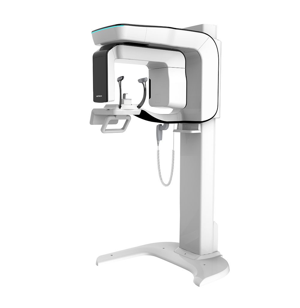
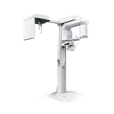
YOUR FIRST PARTNER FOR 3D DIAGNOSIS,
PaX-i3D
| Optimal FOV Sizes for 3D Diagnosis | - Increase your diagnosis and treatment accuracy.
- Multi FOV sizes range from 5x5 to 12x9
|
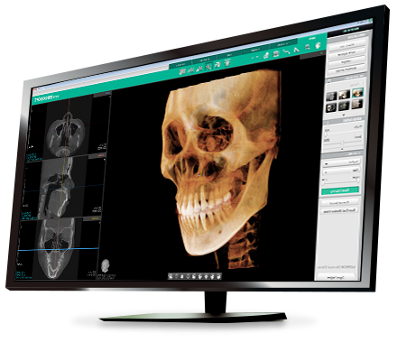
| Optimal FOV Sizes for 3D Diagnosis | - Analyze Ez3D-i images with advanced tools and functions
- Ez3D-i supports effective and efficient communication with your patients
|
| Wide Range of Ceph Modes | - Scan Type: LAT / Full LAT
- One Shot Type: Small / Medium / Large
|
POWERFUL DIAGNOSTIC VALUE WITH 3D IMAGES
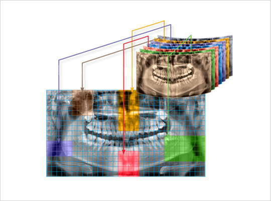
FLEXIBLE 3D IMAGING WITH MULTI FOV SELECTION
PaX-i3D provides 4 multi FOV sizes ranging from 5x5 to 12x9.
By selecting the appropriate FOV size, you can have the optimum image for your diagnostic needs reducing unnecessary X-ray radiation for patients.
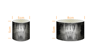  |
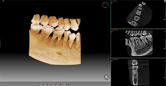
FOV 5X55X5 images are useful for specific area diagnosis with minimum X-ray exposure for patients, It can especially increase the accuracy of endodontic diagnosis by exactly checking the amount or root canals and abnormal root canal shapes such as C-shapes that are difficult to check using 2D X-ray system.
| 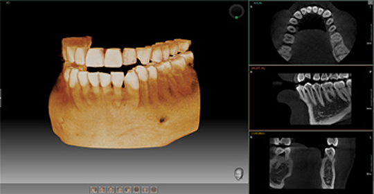
FOV 8X58X5 images can provide more extended oral information on maxillary or mandibular areas. An accurate treatment plan can be established by taking into account the major anatomical structures like mandibular nerve, mental foramen or maxillary sinus.
|
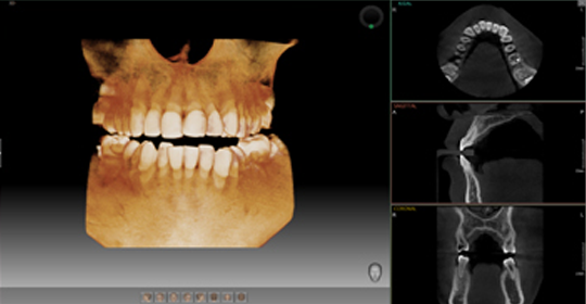
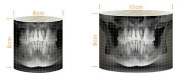
FOV 8X88X8 images enable comprehensive diagnosis and treatment planning including both maxillary and mandibular areas in a single scan. It is useful for complex implant surgery as well /as left or right TMJ diagnosis. |
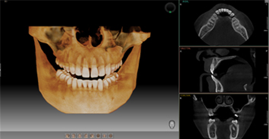
FOV 12X912X9 images can provide the most optimal information for oral diagnosis fully covering both maxillary and mandibular structures including the 3rd molar region in a single scan. It is suitable for most oral surgery cases as well as multiple implant surgery.
|
PROFESSIONAL DIAGNOSTIC VALUE WITH
PANORAMIC IMAGES
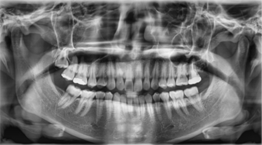
PaX-i3D provides the most precise and high quality panoramic image. Clear and sharp panoramic image brings you better diagnostics. Enhanced details especially in the anterior and dental roots can be viewed. These consistently high quality images will become the new standard of panoramic imaging.
| MAGIC PAN
MAGIC PAN creates a more superb panorama image.
It is acquired through the elimination of distorted and blurred images caused by improper patient positioning (Optional).
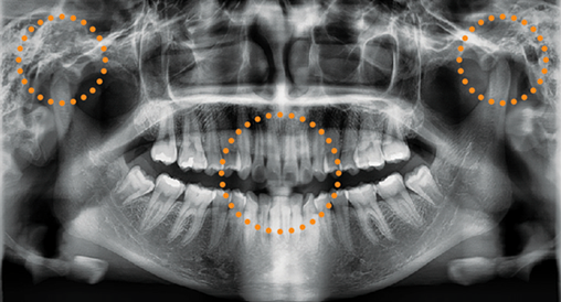 NORMAL PAN
| 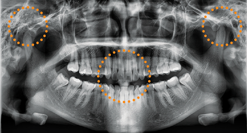 MAGIC PAN
|
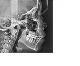
PROFESSIONAL DIAGNOSTIC VALUE WITH CEPHALOMETRIC IMAGES
PaX-i3D Provides optimal images with an EXCLUSIVELY DESIGNED SENSOR FOR CEPHALOMETRIC DIAGNOSIS
SCAN TYPE CEPHALOMETRIC
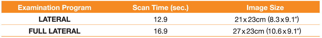
Scan type cephalometric offers two image sizes, LAT and FULL LAT, you can choose one of them based on the purposes of your diagnostic needs
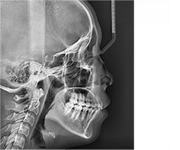
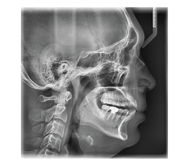 LATERALProvide specialized high quality images to suit orthodontics and maxillofacial surgeries
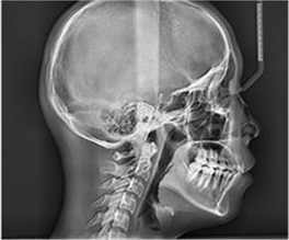
| 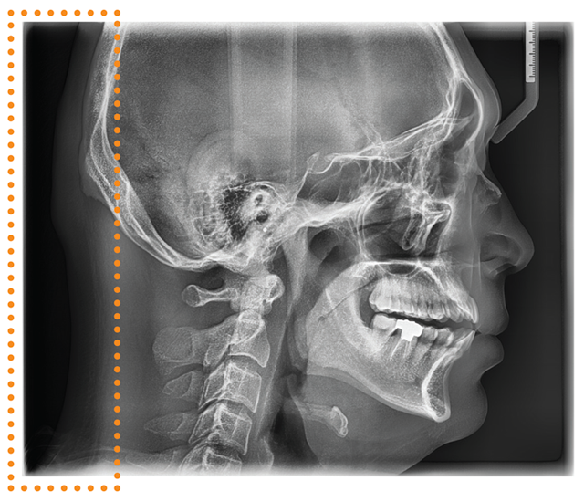 FULL LATERALFull lateral gives 30% larger images and the occipital area of the patient for comprehensive diagnosis. (optional)
|
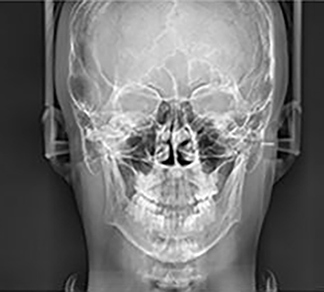
ONESHOT TYPE CEPHALOMETRIC
Three different ceph image sizes reduce unnecessary X-ray dosage and scans the ideal area of cranial anatomy for your diagnosis and treatment planning.
SMALL 20x20cm (8x8")
| MEDIUM 23x35cm (9x10")
| LARGE 30x25cm (12x10")
|
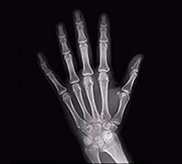
PA
| Carpus
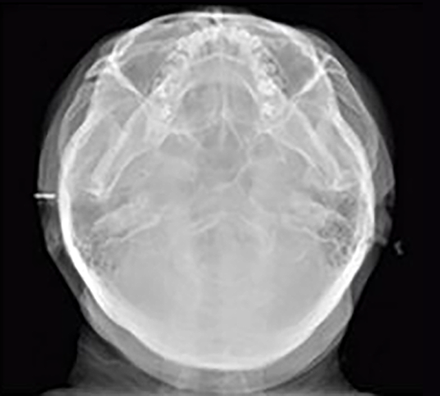
| SMV (Submentovertex)
|
LATERAL
Function | Pano + CBCT + Ceph
|
| CT - FOV Size(cm) | 8X8 : Multi [5X5 / 8X5 / 8X8]
12X9 : Multi [5X5 / 8X5 / 8X8 / 12X9]
|
| Voxel Size | 5X5, 8X5 cm : 0.12mm / 0.2mm,
8X8, 12X9 : 0.2mm / 0.3mm
|
| Scan Time | Pano : HD 13.5 sec (Normal 10.1 sec)
Scan Ceph : 12.9 sec
One Shot Ceph : 0.9 - 1.2 sec
CBCT : Standard 15 sec / High 24 sec
|
Ceph FOV Size
|
21X23cm (8.3"X9.1") [LAT,PA,SMV,Waters View,Carpus]
27X23cm (10.6"X9.1") [Full LAT] | 20X20cm (8"X8") [LAT, PA]
23X25cm (9"X10") [LAT, PA]
30X25cm (12"X10") [LAT, PA, SMV, Waters View, Carpus]
|
|
Gray Scale
| 14 bit
|
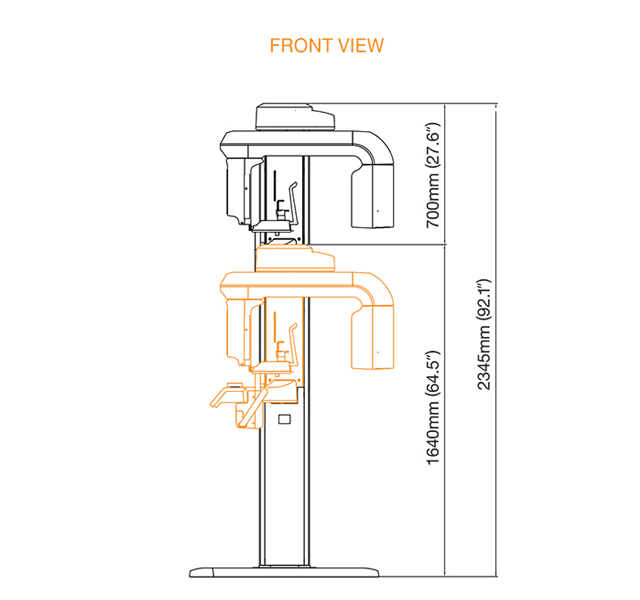
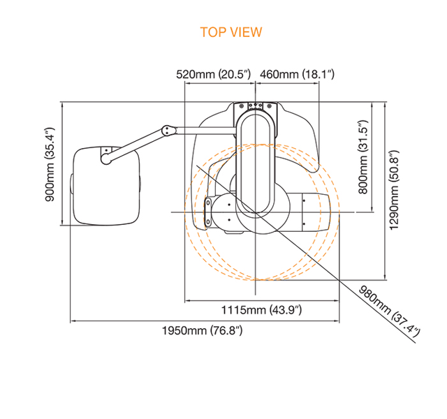
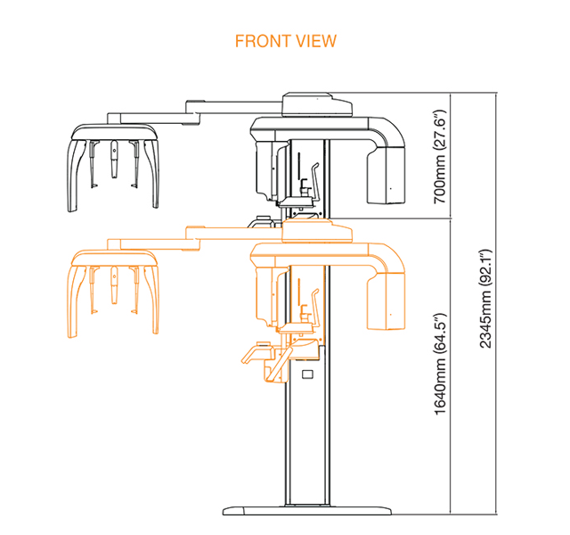
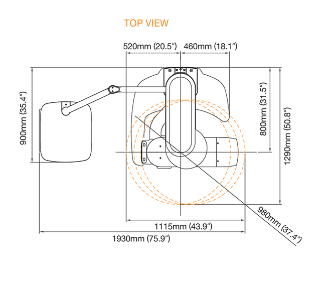
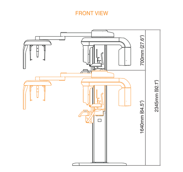
Patient Position
| Standing / Wheel-Chair Accessible
|
Tube Voltage/Current
| 50-90 kVp / 4 - 10 mA
|
The specifications are subject to change without prior notice.
GET QUOTE
REGISTER YOUR VATECH UNIT WITH SCIVISION (SOUTHERN AFRICA)
CBCT COURSES
Cutting Edge Software
 Discovery of new 3D era
|  |
.png)